Del Mar Photonics
Sample quotes for AFM/STM/NSOM
HeroN
Femtosecond NSOM-AFM-STM (Request
a quote)
FEMTOSECOND NSOM
NSOM MoScan-F near-field scanning optical microscope (NSOM) platform
Main Features:
•FleaScan near-field and atomic force microscope unit
•Completely computer controlled xyz- coarse and scan motion
•Optical microscope console with long working distance objectives: simultaneous
probe and sample observation in confocal configuration with submicron
resolution.
•Near-field optical and fluorescence images in photon counting mode
•Near-field optical images in collection and illumination modes
•Transmission and reflection configurations
•Near–field images with lamp illumination (included)
•Near–field images with laser illumination (included)
•Ambient light protection with light-tight box
Configuration:
1. FleaScan near-field and atomic force microscope unit
(a) XY- flat scanner piezo stage, based on a “flea-scan” principle, with 20 mm
central opening
•lateral resolution: better than 100 nm (depends on fiber probe and sample under
investigation)
•maximum xy-travel (computer controlled): 10 mm
•maximum xy-coarse rate: 2 mm/min
•maximum xy-scan: 40 μm x 40 μm
•maximum xy-scan rate: 5 μm/sec
•minimum xy-step: 0.1 nm
•maximum image: 1024 x 1024 pixels
(b) Piezo – inertial z-stage
•z-scanning range – 5 μm
•z- coarse slip-and-stick motion upward or downward, automatically controlled
•maximum z-travel: 9 mm
(c) Fiber probe holder, connected to z-stage and equipped with manually adjusted
xy - micro stage
(d) N.A. = 0.6 optical objective for sample illumination or light collection
2. Optical unit for simultaneous probe and sample observation with long working
distance objectives
•Standard upright optical microscope console with turret and binocular eyepiece
tube mounted to XYZ-manual stage
•20x infinity corrected objective (20.0 mm working distance)
•5x infinity corrected objective (34.0 mm working distance)
•CCD color camera
3. Photon counting PMT, mounted with preamplifier and HV power supply
•Maximum photon counting rate: 10^7 counts/sec
•Dark counts: < 10 cps
•PMT spectral response: 185 - 680 nm
•Adapted to fiber probe (collection mode) or fiber lead (illumination mode) via
light-tight filter holder
4. Filter holder, adapted to PMT unit and to a fiber probe (near-field
collection mode) or a light guide (near field illumination mode)
•Required filters are available optionally.
5. Vibroisolated breadboard and light-tight box for ambient light protection
•Units 1-4 are installed on the breadboard
6. 150 W quartz-halogen illuminator with fiber lead and filters for “cool light”
illumination
7. 1 mW, 532 nm solid-state laser for the sample illumination through a fiber
probe
8. Electronic control unit, containing:
•xy-stage electronics
•z-stage electronics
•Feedback (shear-force) electronics
•Photon counting electronics
•Lock-in amplifier
•Connected to the computer via PCI card
•Power input in the range of 100 – 240 V, 50 – 400 Hz
9. Software
•FleaScan Windows based data acquisition software.
•FemtoScan Windows based image processing software
10. Two years warranty (24 months after installation, not more than 25 months
after date of shipment) for all parts is included on the return to base (RTB)
basis
•MoScan-F optical unit should be installed on an optical or microscope table
(required additionally)
•Pentium III, 500 MHz / 128 Mbite or higher PC with PCI slot and Windows XP is
required additionally
11. One week installation and personnel training is included
Accessories Consumables (included)
1. Near-field probes
•Al-coated optical fiber probes with <100 nm aperture attached to quartz
resonator, 10 pieces
2. Test sample
•100 nm – diameter TransFluoSpheres deposited onto a glass slide
Trestles Opus 3 one-box femtosecond laser including DPSS pump
Trestles 100 femtosecond oscillator
200mW output power at 790nm or other preset wavelength in the range 670-790nm
(indicate required wavelength when ordering)
100 fs pulses
Opus DPSS pump laser, 3 W output power
wavelength 532 nm
beam size 2.0 mm
spatial mode TEM00
M squared < 1.1
power stability < 0.4 % RMS
noise < 0.4% RMS
Near-field Scanning Optical Microscope (NSOM) is a versatile tool for nano-characterization
and nanomanufacturing.
Conventional microscopes have fundamentally limited resolution due to
diffraction, but there is no such restriction for near-field interactions, that
is why near-field microscopy is becoming one of the most important techniques
for nano-science.
Possible applications of this tool are characterization
of photonic nanodevices, bio photonics (investigation of cells, viruses, DNA
molecules), nano-chemistry (chemical reactions control), nanoscale
photolithography (processing of photosensitive polymers).
NSOM delivered femto-second pulses can be used for nanometer-scale surface
topology modification. Temporal resolution provided by femtosecond laser opens
wide range of new possibilities such as: transport dynamics studies of
nanostructured materials, pump-probe experiments, ultra fast coherent and Raman
spectroscopy. Spatial optical resolution of the tool is better than 100 nm and
temporal resolution in the pulse operation mode is better than 100 fs. Tunable
CW operation for spectral measurements is also available, wavelength range in
this case is 710-950 nm.
Advanced Nearfield Scanning Optical Microscopy/Atomic Force Microscopy/Scanning
Probe Microscopy systems (NSOM-AFM-SPM) are used for numerous applications in
materials research, including semiconductors, data storage, electronic
materials, solar cells, polymers, catalysts, life sciences and nano-sciences.
NSOM-AFM-SPM is a well-established method for ultra-high nano-scale spatial
resolution surface imaging and the characterization of surfaces and interfaces
down to atomic dimensions.
Request a quote
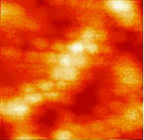
Fluorescence image of 100 nm - diameter TransFluoSpheres,
received under excitation at 532 nm and detection around 600 nm.
|
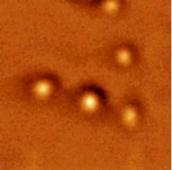
Near-field optical image of 250 nm - diameter gold beads, deposited onto
a glass slide.
Image size: 2 mm x 2 m |
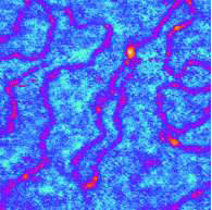
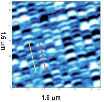
AFM (topography) image of DNA (<3 nm thickness),
deposited onto a glass slide |
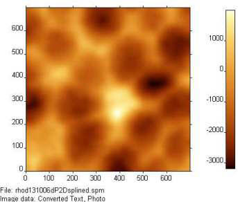
Near-field optical image of 100 nm - diameter polystyrene beads,
deposited onto a glass slide |
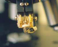
Standard 100kHz fiber probe and fiber micro objective for the reflection
mode operation. |
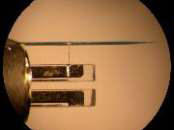
32 kHz custom nanofiber probe
|
NSOM Applications
Photonic Crystal Nanocavities for
Efficient Light Confinement and Emission
pdf
Photonics Interconnects
Fabrication and integration of VLSI micro/nano-photonic circuit board
Plasmonics: Merging Photonics and Electronics at Nanoscale Dimensions
Femtosecond Near-field Scanning Optical Microscope NSOM investigations of pulse
multiwave mixing in Semiconductor Optical Amplifiers
Femtosecond lasers recommended for use with NSOM
Ti:Sapphire lasers
Trestles femtosecond Ti:Sapphire laser
Trestles Finesse femtosecond
Ti:Sapphire laser with integrated DPSS pump laser
Teahupoo Rider femtosecond amplified
Ti:Sapphire laser
Cr:Forsterite lasers
Mavericks femtosecond
Cr:Forsterite laser
Er-based lasers
Tamarack femtosecond fiber laser (Er-doped
fiber)
Buccaneer femtosecond OA fiber laser (Er-doped
fiber) and SHG
Cannon Ultra-broadband light source
Yb-based lasers
Tourmaline femtosecond Yt-doped fiber laser
Tourmaline Yb-SS400 Ytterbium-doped Femtosecond Solid-State Laser
Tourmaline Yb-ULRepRate-07 Yb-based high-energy fiber laser system kit
Cr:ZnSe lasers
Chata femtosecond Cr:ZnSe laser (2.5 micron) coming soon
Del Mar Photonics nano-imaging gallery
High resolution MFM image of Seagate Barracuda 750Gb Hard Drive, ST3750640AS.
130 nm Ag nanoparticles immobilized on the metal surface, 3.6x3.6 um scan
Magnetic structure of surface domains in Yttrium Iron Garnet (YIG) film
Atomic resolution on HOPG obtained with the 100 micron scanner
NSOM Fluorescence image of 100 nm - diameter TransFluoSpheres
Near-field optical image of 250 nm - diameter gold beads, deposited onto a glass
slide
AFM (topography) image of DNA (<3 nm thickness),
deposited onto a glass slide
Near-field optical image of 100 nm - diameter polystyrene beads, deposited onto
a glass slide
Send
us your sample for nano-characterization!!!
Related Del Mar Photonics Products:
AFM HERON
Near-field
Scanning Optical Microscope (NSOM)
Femtosecond nanophotonics
Femtosecond NSOM
SPIE Photonics West 2009 product announcement
Conventional microscopes have fundamentally limited resolution due to
diffraction, but there is no such restriction for near-field interactions, that
is why near-field microscopy is becoming important nano-science technique.
Possible applications of this tool are characterization of photonic nanodevices,
bio photonics (investigation of cells, viruses, DNA molecules), nano-chemistry
(chemical reactions control), nanoscale photolithography (processing of
photosensitive polymers). NSOM delivered femto-second pulses can be used for
nanometer-scale surface topology modification. Temporal resolution provided by
femtosecond laser opens wide range of new possibilities such as: transport
dynamics studies of nanostructured materials, pump-probe experiments, ultra fast
coherent and Raman spectroscopy. Spatial optical resolution is better than 100
nm and temporal resolution in the pulse operation mode is better than 100 fs.
Tunable CW operation for spectral measurements is also available.
Request a quote